FFR and EFR
with AI
from a single
CT scan.
VCAST is an AI-powered platform that derives functional coronary data – fractional flow reserve, energy flow reserve, and plaque burden – from standard CTCA. No pressure wire. No stress agent. No additional acquisition.
Three reasons
CAD is still diagnosed in fragments.
Invasive.
FFR remains the gold standard – but measuring it requires a pressure wire inside the coronary artery. Only 6% of eligible patients receive it. The procedure carries cost, cathlab access, and procedural risk.
Incomplete.
CT angiography shows anatomy – lumen width, plaque location. It does not show flow. A 70% stenosis on imaging may be haemodynamically insignificant, or a 50% stenosis may be the one causing ischaemia. Anatomy alone cannot decide.
Inefficient.
Nuclear stress tests, echo stress, and cathlab FFR add days or weeks to the diagnostic pathway. Each step occupies capital equipment, specialist time, and increases patient exposure. The pathway fragments across departments.
A virtual
stress test, built
from a single CT.
VCAST is a patented, AI-based medical system that performs non‑invasive, quantitative analysis of standard CTCA data. It produces fractional flow reserve (FFR), energy flow reserve (EFR), and plaque burden – the functional data normally reserved for the catheterisation laboratory – without pressure wires, stress agents, or specialist intervention.
The only input required is a standard CT acquisition. No modified protocol. No additional equipment. Any scanner, any vendor, any imaging protocol.
A standard non-invasive CT scan
No extra acquisition
AI segmentation, verification + CFD simulation.
Everything in cloud
FFR, EFR, plaque burden – 3D models
With access on every mobile device
Any CT manufacturer, any protocol
Fully agnostic
Six stops,
one path,
every patient.
From acquisition to interactive report… with radiologists in the loop
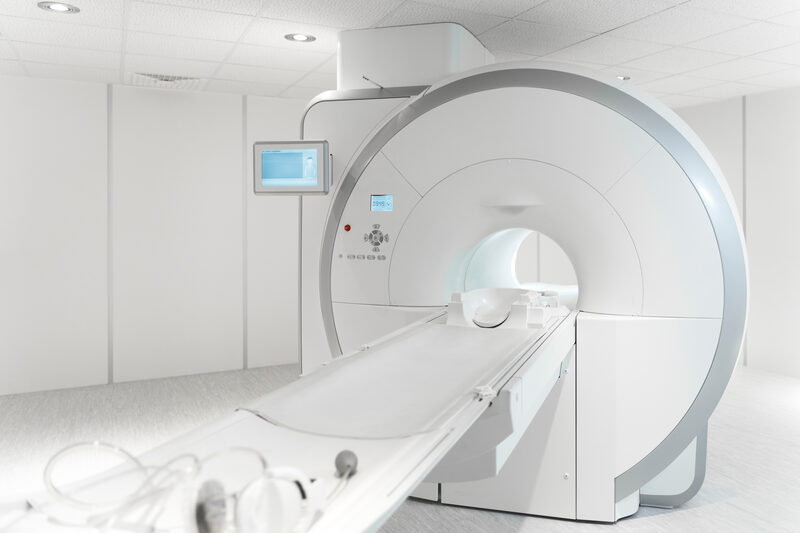
Computed tomography
A standard non-invasive CT – the only input VCAST requires. Independent of scanner type or imaging protocol.
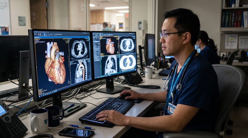
DICOM ingestion
Upload directly or stream from your PACS via the VCAST API. Metadata extracted automatically.
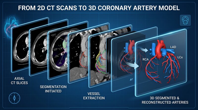
AI segmentation
A proprietary cloud algorithm reconstructs a full 3D coronary model from 2D slices in minutes.

Expert verification
A team of radiologists reviews and signs off on every segmentation. AI does the lift, humans approve.
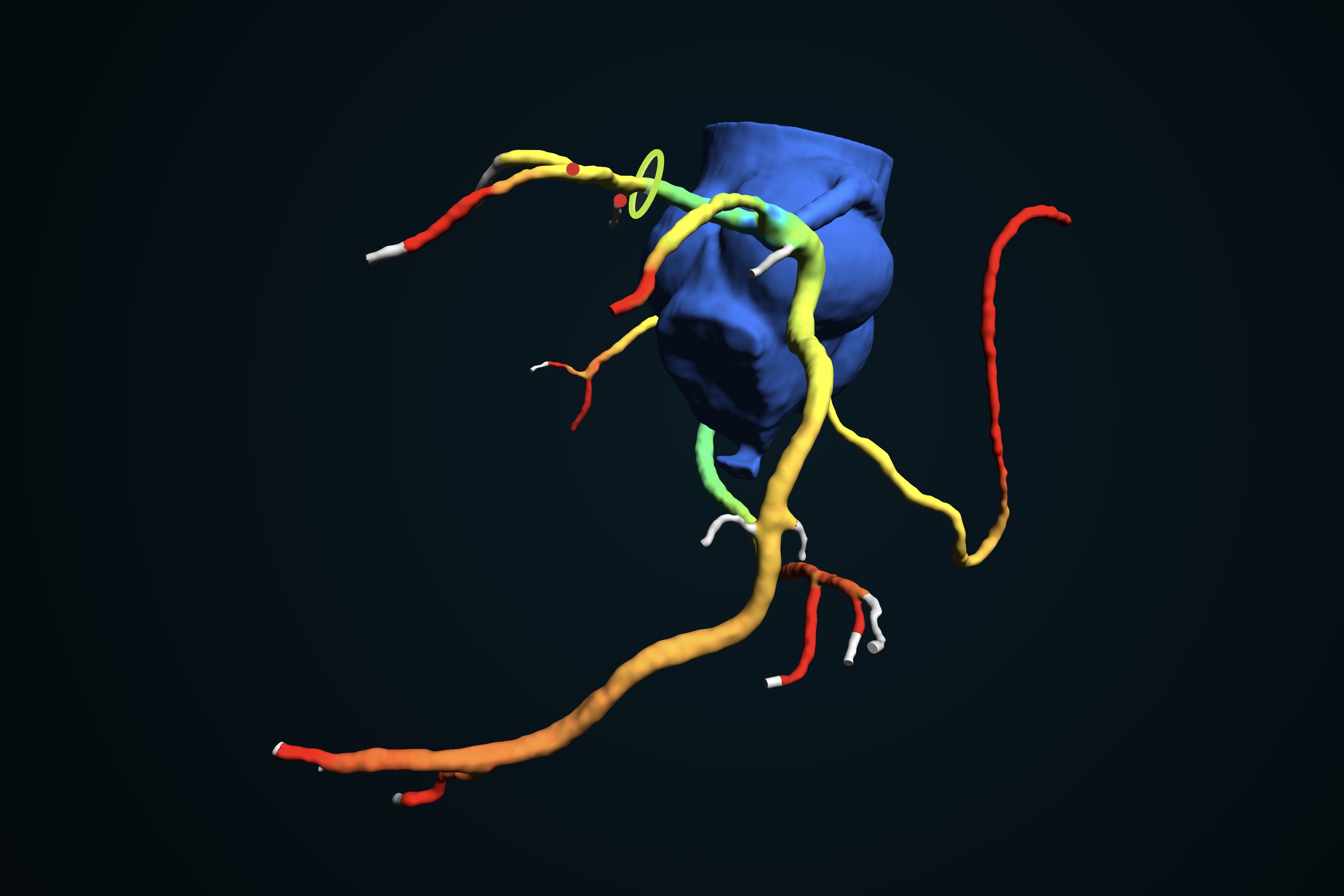
CFD simulation
Computational fluid dynamics simulate pressure, velocity and FFR across every named segment.

Interactive report
A full report, with embedded 3D models, reaches the physician through QR or web – wherever they read.
What clinicians and partners gain.

Non-invasive
No catheters, no stress agents. The CT they already had is enough.

100% Cloud
Auto-scales with caseload. No on-premise hardware. No additional infrastructure. Simple, powerful and intuitive. It only takes 5 minutes to learn and start working.

Decision-grade outputs
FFR, EFR, stenosis grade – in one colour-coded viewer available on any mobile device. The data normally available only from the cathlab.

AI + radiologist QA
AI performs segmentation autonomously. A radiologist reviews and signs off every case before results are released.

PACS-native
Stream DICOMs directly from your PACS – no downloads, no manual file transfer. Compatible with any vendor and any imaging protocol.

CE-marked, reimbursement guidance
CE-marked under MDR. Coding and reimbursement guidance available for target markets. Contact us for country-specific information.

Backed by
peer-reviewed
science.
VCAST is supported by published research, multi-centre validation, and reimbursement guidelines in our target markets. We share the full evidence dossier on request.
Diagnostic accuracy of CT-derived FFR using AI-based 3D segmentation: a multi-centre study.
Concordance between VCAST-derived FFR and invasive measurement across vendor-heterogeneous CT acquisitions.
Triage of suspected CAD using virtual stress testing — operational impact at a tertiary centre.
Use of CT-derived hemodynamic indices in stable chest pain pathways (Class IIa).
The full white paper – methodology, validation cohorts, regulatory file.
Project
Kardiobeat.CT

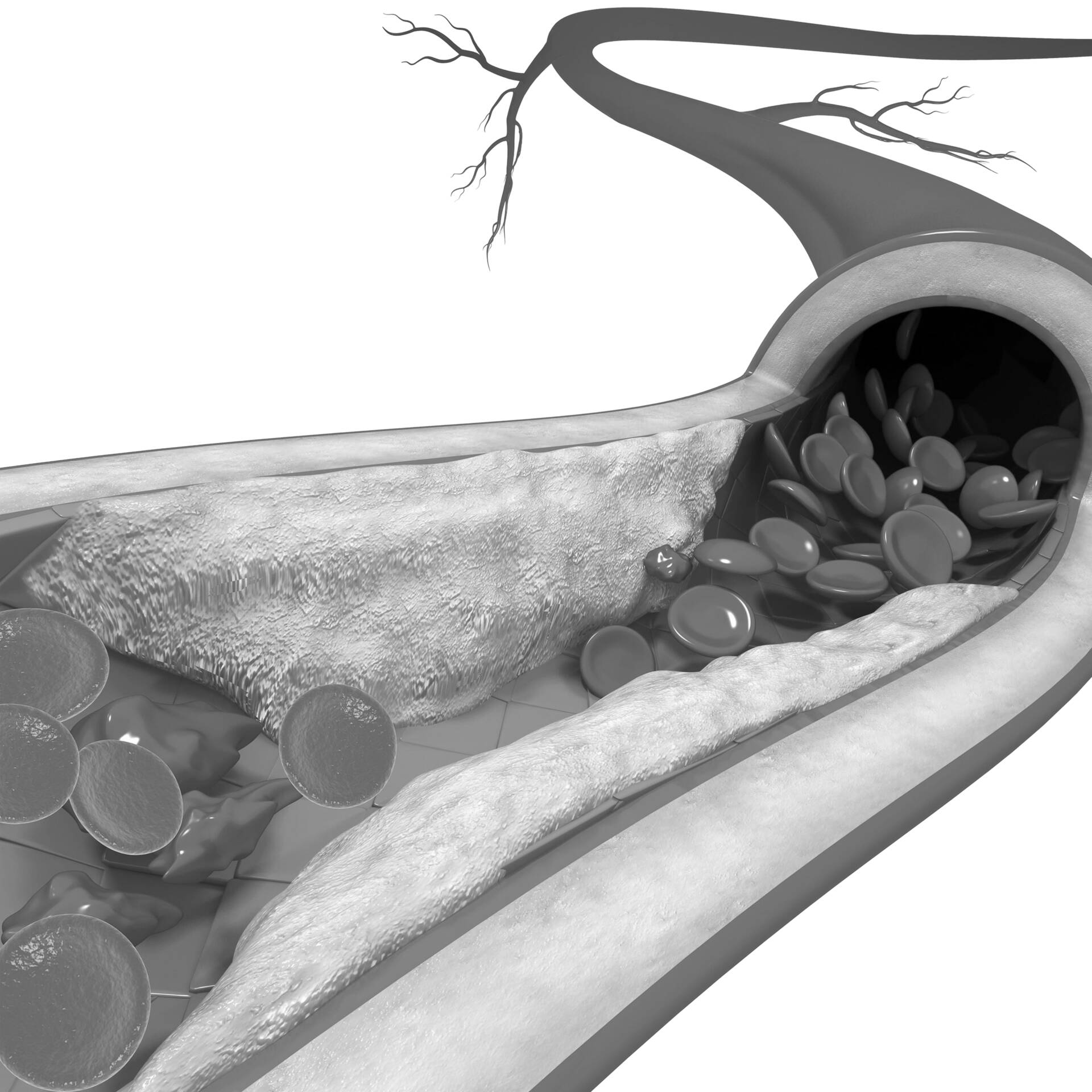
The aim is to create an innovative diagnostic tool for the automatic detection and assessment of atherosclerotic plaques – a solution currently unavailable on the market. Supporting the project through PARP financing will enable the creation of an innovative product that can be a pioneer on the global market.
- •full characteristics of the plaque
- •classification of plaque according to the degree of calcification
- •distance from the ostium
- •full values of the plaque volume
What physicians
get.
The VCAST report delivers per-vessel FFR, EFR in a colour-coded 3D viewer – accessible via browser or QR code from the printed report. No dedicated workstation required.

The first time I saw VCAST results next to my invasive FFR measurement, the two were within rounding error. It changed how I triage stable chest pain.
Dr. [Cardiologist]
Interventional cardiologist · Tertiary cardiac centre
The report lands on the workstation in fifteen minutes — including the interactive viewer the clinician actually opens. That alone removed our second-look bottleneck.
Dr. [Radiologist]
Head of cardiac imaging · University hospital
The report lands on the workstation in fifteen minutes — including the interactive viewer the clinician actually opens. That alone removed our second-look bottleneck.
Dr. [Radiologist]
Head of cardiac imaging · University hospital
~15 minutes
5k+
0.01
38
See our CT
in VCAST.
Book a 30-minute call with us.
Prepare a de-identified CT case — we'll process it
for you so you can see the result
A live analysis on demo CT
Full 3D viewer walkthrough
Integration & reimbursement Q&A
Evidence dossier bundle